The human eye
It is available to us around the clock with its numerous capabilities giving us, among other things, spacial awareness. It is also responsible for differentiating colours, shapes, speeds, movement as well as distances and, last but not least, recognising people and complex situations.
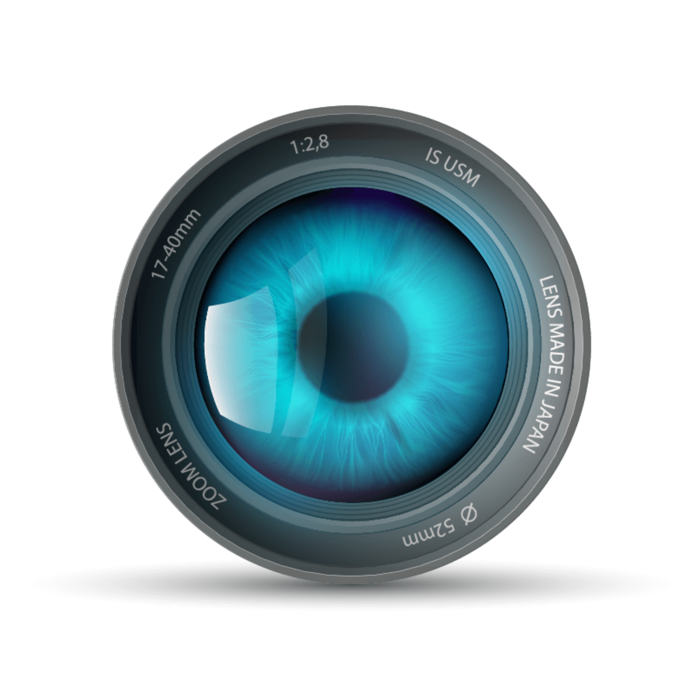
Comparison with a camera
The light reaches the sensor or film/ the retina, via the camera’s lens/ the eye’s lens, which uses autofocus/ accommodation to set the right distance, and the aperture/ the pupil, to regulate the amount of incoming light.
An ingenious process which mother nature thought up and implemented all by herself.
Many complex tasks
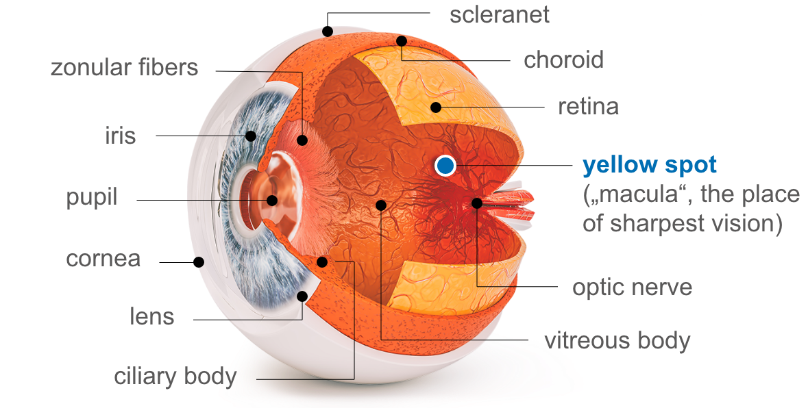
The light enters the eye via the clear and transparent cornea at the foremost part of the eye. It is therefore like an optical window. Its high level of sensitivity causes an eyelid closure reflex in response to even the smallest touch. The cornea also provides the eyeball with the necessary stability in the frontal region. The white sclera provides the necessary stability and shape to the side and back of the eye. The conjunctiva is directly adjacent to the cornea. It covers the sclera and the inside of the eyelids. The conjunctiva firstly protects against germs and secondly ensures that the eyeball is able to move in all directions. To ensure that the cornea does not dry out and cloud over, it is continuously and automatically moistened with lacrimal fluid.
The light then enters through the pupil which is encircled by the annular iris, its pigmentation being responsible for the eye colour. The pupil regulates the amount of light entering the eye by changing its size. In light surroundings, the pupil constricts (state of myosis) and in dark surroundings, it widens (state of mydriasis). This process is carried out by muscles in the iris. The iris at the same time separates the anterior and posterior chambers of the eye.
Once the light from outside has penetrated the pupil, it reaches the retina via the eye’s lens (Lens cristallina) and the vitreous body (Corpus vitreum), which is composed of a gelatinous mass and gives the eye stability. The eye’s lens collects the light which falls on it. It reaches the retina as a bundle and provides a clear image. The eye’s lens is affixed by way of numerous small ligaments (zonules) on the ciliary body and by tensing the muscles there it can change in such a way as to allow near and distance vision. This process is described as accommodation.
The retina’s role is to sense the light via photoreceptors by transforming light pulses into nerve signals, and then direct these to the brain via the optic nerve papilla, where all optic nerves meet. The brain is where vision actually takes place.
The macula lutea
This is located in the centre of the retina next to the junction of the optic nerve. The centre of the macula has a small depression, which has the highest density of light receptors – the point of sharpest vision.
While focusing on an object with the eye, the angle of sight is readjusted constantly by automatic tiny rotational movements of the eyeball, so that the object is precisely replicated on this central depression of the macula lutea. Pathological changes in the macula (e.g. as part of age-related macular degeneration) therefore compromise the visual acuity.

The name macula lutea translates as “yellow spot” and is so called because of the high concentration of the pigment of lutein. Lutein is an important ingredient in the AREDS2 formulation – although not sufficient alone. It is precisely this clinically proven combination which is contained within all PROMACULA® products. Here the lutein has an antioxidative effect and protects the macula against harmful environmental influences.
The choroid
The choroid is situated between the outer sclera and the inner retina. It consists of a very thick network of blood vessels. This network is responsible for nourishing the outer retinal layers.
The vascular tunic (Uvea)
The uvea is a construct, an interaction between
- the annular iris which, by changing its size, regulates the amount of light entering the eye;
- the ciliary body behind the eye’s lens on the sclera, which uses its muscles to take on the role of accommodation (focusing) for near and distance vision, as well as the production of tears;
- and the choroid which is responsible for nourishing the outer retinal layers.
Where tears are created
Above the eye, slightly to the side in the eye socket is where the lacrimal gland is located. This is where tears are produced and which are then distributed around the eyeball by the eyelids. From there, they are transported into the nose via the lacrimal ducts which start in the inner corner of the eye.
What precorneal film consists of
The precorneal film consists of three layers. The mucosal layer, or mucin, lies directly on the cornea. It’s role is to ensure that the precorneal film remains adhered to the surface of the eye.
The middle layer of the precorneal film largely consists of water. This layer contains many dissolved substances such as antibodies against infection.
The very outside of the corneal film is covered with a fine lipid layer. This involves a film of oil which stops the corneal film from evaporating. This oily layer is formed by the sebaceous glands in the eye lids (Meibomian glands).
Dry eyes
If there is a reduction in the quantity of lacrimal fluid (e.g. due to a lack of omega-3 fatty acids which support the formation of lacrimal fluid) and/or a changed composition of the lacrimal fluid, this will result in a dysfunction in the lubrication of the eye’s surface. These changes result in the eye no longer being lubricated.
The patient experiences dry eye (Sicca syndrome or Sjogren’s syndrome) as burning, reddening, itching and a foreign body sensation and it can subsequently promote the development of AMD.
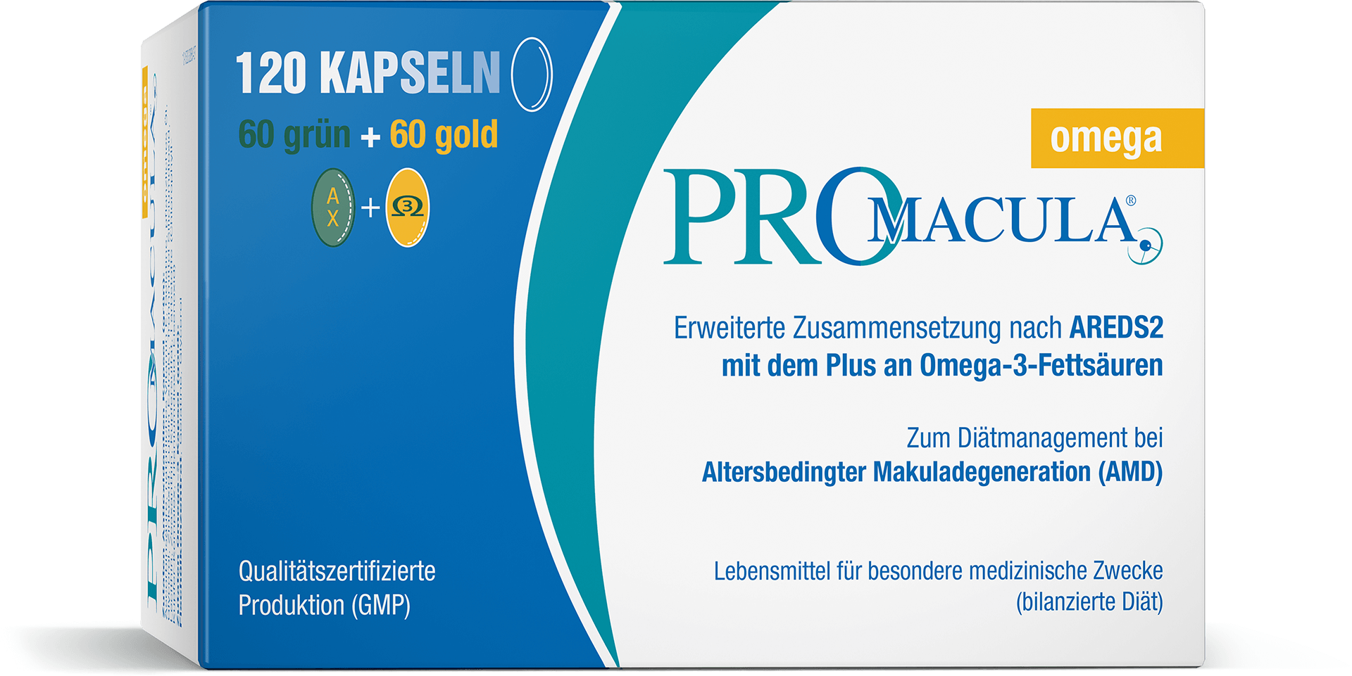
PROMACULA® omega
supplements the well-proven AREDS2 formulation with high-quality omega-3 fatty acids and is therefore the ideal product for treating AMD when the disease is accompanied by “dry eyes”.
Functioning of the iridocorneal angle and the eyelids
The iridocorneal angle is situated at the point where the cornea meets the sclera. Here there is a small channel from which the constantly produced aqueous fluid drains out of the eye.
The eyelids are first and foremost there to protect the eyes. The lashes keep out foreign bodies and the previously mentioned eyelid closure reflex works to close the eyes as quickly as possible when at risk. The eyelids also act to lubricate the cornea, stopping it from drying out.
What makes the eyes move?
In all there are six different eye muscles responsible for eye movement:
four straight and two oblique muscles.
In short: our eyes are a masterpiece of nature, but they are also very sensitive