What is wet AMD?
This form of macular degeneration is characterised by the growth of new but abnormal blood vessels coming from the choroid membrane, which grow into and under the retina. These new vessels are permeable so fluids leak out of them. This results in swelling on the retina, and it’s not uncommon for bleeding to occur.
Preventing wet AMD
Since wet macular degeneration generally represents a negative advancement of dry AMD, it is important that the dry form is detected early.
A healthy diet with sufficient vitamins and trace elements (zinc is particularly important) over as many years as possible is considered beneficial. Smoking should be avoided at all costs. But if there is already a diagnosis of advanced AMD, a healthy diet alone is no longer sufficient, as very high doses will now be needed.
Ask your eye specialist about PROMACULA® as a preventative treatment against wet AMD.
The AREDS2 formulation of PROMACULA® protects your eyesight.
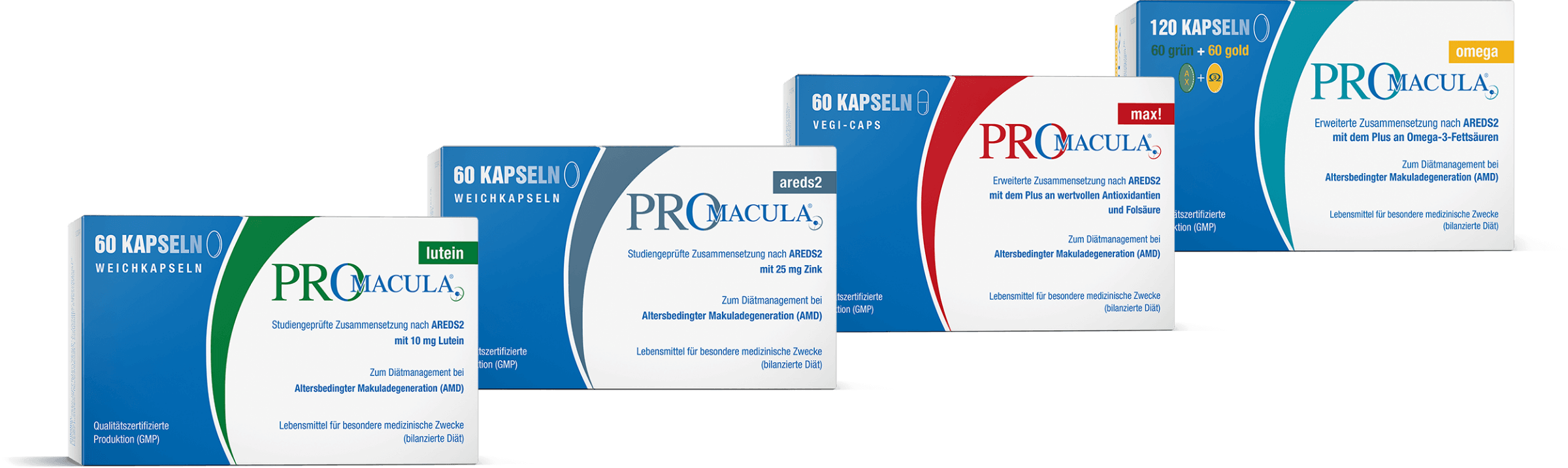
PROMACULA® can provide preventative treatment against aged-related, wet AMD. The preparation was developed based on the results of the ARED studies (Age-related Eye Diseases).
Based on this, a research team at the US National Eye Institute was able to prove that a specific combination of different nutrients were crucial in fortifying the eye’s protective system, thus slowing down the progression of AMD.
If advanced AMD disease has already been diagnosed, it is essential that appropriate action is taken to slow down further progression of the disease. For example, if one eye is already affected by wet AMD, taking PROMACULA® can reduce the changes of the other eye also developing wet AMD. Patients with advanced stages of dry AMD have an increased risk of suffering from loss of sight and developing the wet form of AMD. The results of the AREDS2 study have shown that this risk can be considerably reduced by taking an AREDS2 compliant preparation (such as PROMACULA®).
Diagnosis of wet AMD
The diagnostic assessment starts with carrying out a basic examination to determine visual acuity. Then the cornea, sclera, lens, ocular fundus and the vitreous body of the eye are examined using the slit lamp. Then follows an ophthalmoscopy, also known as a fundoscopy. The visible parts of the eye, mainly the retina and the blood vessels which supply it, are also examined.
Fluorescence angiography, a specific examination of the whole ocular fundus, examines the bloods vessels in the retina and under the retinal pigment epithelium in the choroid membrane.
Using angiography, an examination using dye, allows changes in the blood vessels to be shown. Dye injected into the veins of the arm reaches the blood vessels in the eyes through the blood circulation, making the vessels easier to see. A series of photographs is then taken over a period of approx. 10 minutes.
A high resolution optical coherence tomography (OCT) is often carried out, which is a non-invasive scan of the retina using a laser beam. This allows a cross-section of the retina to be created based on up to 50,000 measurements per second. If a wet AMD is present, it will show the fluid accumulations in and under the retina. If there are no accumulations of fluid, but deposits under the retina and a thinning of the retinal structure, then this indicates dry AMD. This examination is available privately, paid for by the patient themselves.
The Amsler grid test is recommended for self-testing. If the black grid is not perceived as a continuous, regular and straight image, a check-up should be arranged to find out whether it is evidence of AMD. [Click here to go to the self-test]
If AMD is detected promptly, targeted treatment can be effective at counteracting it.
What symptoms are associated with wet AMD?
It starts with difficulties with reading, a clear sign being the distorted vision of straight lines or rows of letters. If wet AMD continues to progress, a black or grey spot will appear in the centre of the field of vision. Sight around this spot usually remains intact. [More information]
In advanced stages, familiar faces are no longer recognised in the street. Reading and writing, as well as manual work and many other everyday activities such as driving become a problem and at some point become impossible. In the advanced stages, it becomes virtually impossible to continue living an active, independent life with social, cultural and sporting activities.
Take prompt action: eyesight is quality of life.
Treatment options for wet AMD
Treatment with VEGF inhibitor
The aim of this treatment is firstly to reverse the unwanted new formation of vessels and, secondly, to prevent the formation of new vessel growth.
A VEGF inhibitor is injected directly into the diseased eye. If both eyes suffer from wet AMD, the VEGF inhibitors are injected into both eyes. Unfortunately, the anti-VEGF therapies do not primarily enable permanent deactivation of new vessel formations. This is why treatment has to be repeated in regular intervals and can in some cases continue for several years.
What is the aim of this injection treatment (anti-VEGF treatment)?
It aims to inhibit the release of the VEGF (vascular endothelial growth factor) hormone because VEGF is the cause of unwanted vascular growth (neovascularisation) as well as the greater permeability of the vessel walls. This causes the dreaded haemorrhaging, or release of fluid, into the eye. The aim is to transform the wet AMD into the dry form.
It should be noted that anti-VEGF therapy has replaced the PDT method, a photodynamic therapy, as the most effective method in conventional medicine to date.
OCT (Optical Coherence Tomography) of the retina is carried out for follow-ups and to check the success of the treatment.
The target continues to be to minimise the permanent structural damage caused by the degeneration of the macula. This is why the injection treatment of wet AMD is performed with the aim of stabilising and limiting the active phase.
To ensure its success, the aim of reducing the retinal oedema and inhibiting further neovascularisation is to minimise the risk of further progressive macular degeneration.
Pre-treatment for a VEGF inhibitor therapy
First, fluorescence angiography of the retinal vessels is performed. This assesses whether this treatment can be successful. Before the VEGF inhibitors are injected in a sterile outpatient room, the conjunctival sac is flushed out. Then the entire eye area is disinfected and the lashes are taped out of the way using film. Then comes the local anaesthetic in drop form.
The injection is then performed intraocularly, that is directly into the rear section of the eye, the vitreous body. This procedure is performed in the pars plana, so just around 4 mm from the cornea. These intravitreal injections are used so that a high concentration of the active ingredient can reach precisely the place where its effect should be deployed.
Currently, a customised procedure, PRN, is used. Over the first three months, the VEGF inhibitors are injected once each time. Then the previous and further treatment success is assessed in a comprehensive examination. If the prognosis is positive, the treatment is continued as before. If not, it will be discontinued. If it is continued, another assessment is carried out after a month in the hope that the wet AMD has become dry.
The treat-and-extend method of treating wet macular degeneration
The injection of the VEGF inhibitors is carried out monthly until the wet AMD has transitioned into a dry form. Then, further treatment is carried out, checked at longer intervals and the injection time intervals thus gradually extended until the optimal rhythm is found so that the macula remains dry.
According to a study at the Casey Eye Institute (CEI) in America, after approximately a year of continuous treatment, a stabilising effect is seen in around 70 % of patients.
Magnifying visual aids to provide relief at work and in everyday life
In line with the wet AMD stage, reading can usually be improved with magnifying visual aids. This includes not only reading glasses but also special magnifiers or a reading device.
Reading or magnifying glasses can help to improve reading.

Can wet macular degeneration be cured?
The currently available treatments do not offer a cure, because, unfortunately, they do not primarily enable permanent deactivation of neovascularisation. This is why it’s all the more important when receiving your diagnosis, to discuss the prompt starting of regular PROMACULA® (100% AREDS2 compliant) with your treating doctor.
This can reduce the risk of your AMD progressing.